- Source
- Escherichia coli
- Molecular Weight
- Approximately 24.8 kDa, a disulfide-linked homodimeric protein containing two 110 amino acid residues polypeptide (B chain).
- Sequence
- MSLGSLTIAE PAMIAECKTR TEVFEISRRL IDRTNANFLV WPPCVEVQRC SGCCNNRNVQ CRPTQVQLRP VQVRKIEIVR KKPIFKKATV TLEDHLACKC ETVAAARPVT
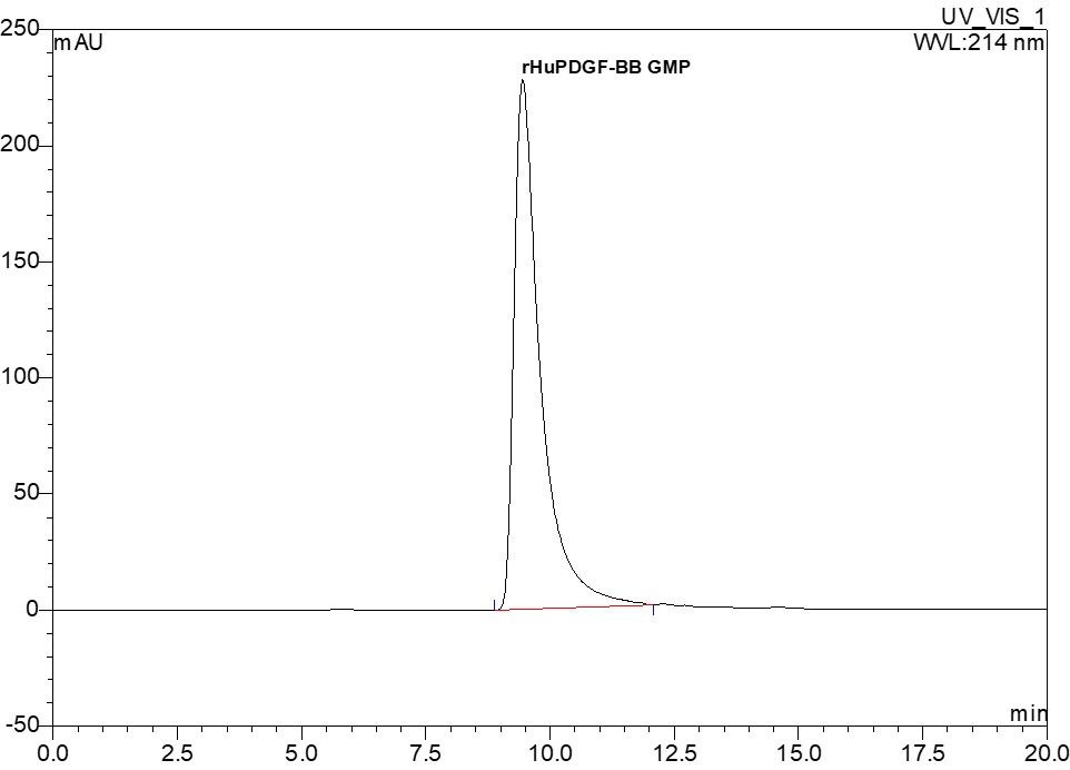
- Purity
- > 98% by SDS-PAGE and HPLC analyses.
- Biological Activity
- Measured in a cell proliferation assay using murine Balb/c 3T3 cells. The specific activity of recombinant human PDGF-BB is > 3.3 × 10⁵IU /mg, which is calibrated against the human PDGF-BB WHO International Standard (NIBSC code: 94/728).
- Physical Appearance
- Sterile filtered white lyophilized (freeze-dried) powder.
- Formulation
- Lyophilized from a 0.2 µm filtered concentrated solution in PBS, pH 7.4.
- Endotoxin
- Less than 0.01 EU/μg of rHuPDGF-BB GMP as determined by LAL method.
- Sterility
- Negative.
- Mycoplasma
- Negative.
- Host Cell Protein
- Less than 0.05% when tested by ELISA.
- Host Cell DNA
- Less than 20 ng/mg when tested by qPCR.
- In Vitro Virus Assay
- Negative.
- Reconstitution
- Prior to opening, it is recommended to centrifuge the vial briefly to bring the contents down the bottom. Reconstitute in sterile distilled water or aqueous buffer containing 0.1% BSA to a concentration of 0.1-1.0 mg/mL. If animal-origin-free condition is expected in your product, then sterile distilled water is recommended. Stock solutions should be apportioned into working aliquots and stored at ≤ -20 °C. Further dilutions should be made in appropriate buffered solutions.
- Shipping
- The product is shipped with polar packs. Upon receipt, store it immediately at the temperature recommended below.
- Stability & Storage
- Use a manual defrost freezer and avoid repeated freeze-thaw cycles.
- A minimum of 12 months from date of receipt, when stored at ≤ -20 °C as supplied.
- 1 month, 2 to 8 °C under sterile conditions after reconstitution.
- 3 months, -20 to -70 °C under sterile conditions after reconstitution.
- Refer to lot-specific CoA for the Expiry Date.
- Usage
- This material is offered by Shanghai PrimeGene Bio-Tech for research, laboratory, or further evaluation purposes. NOT FOR HUMAN USE.
- Quality Statement
- The manufacturing and testing of these products comply with ICH Q7 guidelines.
- Background
- Platelet-derived growth factor (PDGF) presenting in serum but absent from plasma was first discovered in animal study by Lynch and co-workers in the late 1980s. It is a disulfide-linked dimer consisting of two peptides-chain A and chain B. PDGF has three subforms: PDGF-AA, PDGF-BB, PDGF-AB. It is involved in a number of biological processes, including hyperplasia, embryonic neuron development, chemotaxis, and respiratory tubule epithelial cell development. The function of PDGF is mediated by two receptors (PDGFR-α and PDGFR-β).


 COA申请
COA申请
 首页
首页
 Product Datasheet
 MSDS
MSDS
 筛选产品
筛选产品