- Synonyms
- BSF-2, CDF, Hybridoma growth factor, IFN-beta-2.
- Source
- Escherichia coli.
- Molecular Weight
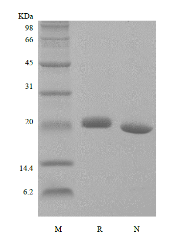
- Approximately 20.8 kDa, a single non-glycosylated polypeptide chain containing 183 amino acids.
- AA Sequence
- VPPGEDSKDV AAPHRQPLTS SERIDKQIRY ILDGISALRK ETCNKSNMCE SSKEALAENN LNLPKMAEKD GCFQSGFNEE TCLVKIITGL LEFEVYLEYL QNRFESSEEQ ARAVQMSTKV LIQFLQKKAK NLDAITTPDP TTNASLLTKL QAQNQWLQDM TTHLILRSFK EFLQSSLRAL RQM
- Purity
- > 96 % by SDS-PAGE and HPLC analyses.
- Biological Activity
- Assay #1: Fully biologically active when compared to standard. The ED50 as determined by a cell proliferation assay using IL-6-dependent murine 7TD1 cells is less than 0.1 ng/ml, corresponding to a specific activity of > 1.0 × 10⁷ IU/mg.
Assay #2: Fully biologically active when compared to standard. The ED50 as determined by a cell proliferation assay using IL-6-dependent murine T1165 cells is less than 0.8 ng/ml, corresponding to a specific activity of > 1.25 × 10⁶ IU/mg.
- Physical Appearance
- Sterile Filtered White lyophilized (freeze-dried) powder.
- Formulation
- Lyophilized from a 0.2 µm filtered concentrated solution in PBS, pH 7.4.
- Endotoxin
- Less than 1.0 EU/µg of rHuIL-6 as determined by LAL method.
- Reconstitution
- We recommend that this vial be briefly centrifuged prior to opening to bring the contents to the bottom. Reconstitute in sterile distilled water or aqueous buffer containing 0.1 % BSA to a concentration of 0.1-1.0 mg/mL. Stock solutions should be apportioned into working aliquots and stored at ≤ -20 °C. Further dilutions should be made in appropriate buffered solutions.
- Stability & Storage
- Use a manual defrost freezer and avoid repeated freeze-thaw cycles.
- 12 months from date of receipt, -20 to -70 °C as supplied.
- 1 month, 2 to 8 °C under sterile conditions after reconstitution.
- 3 months, -20 to -70 °C under sterile conditions after reconstitution.
- Usage
- This material is offered by Shanghai PrimeGene Bio-Tech for research, laboratory or further evaluation purposes. NOT FOR HUMAN USE.
- SDS-PAGE

- Reference
- 1. Ferguson-Smith AC, Chen YF, Newman MS, et al. 1988. Genomics. 2:203-8.
2. van der Poll T, Keogh CV, Guirao X, et al. 1997. J Infect Dis. 176:439-44.
3. Ming JE, Cernetti C, Steinman RM, et al. 1989. J Mol Cell Immunol. 4:203-11; discussion 211-2.
4. Bastard JP, Jardel C, Delattre J, et al. 1999. Circulation. 99:2221-2.
5. Heinrich PC, Behrmann I, Muller-Newen G, et al. 1998. Biochem J. 334 ( Pt 2):297-314.
- Background
- Interleukin-6 (IL-6) is an interleukin that in humans is encoded by the IL-6 gene and acts as both a pro-inflammatory and anti-inflammatory cytokine. It is secreted by T cells and macrophages to stimulate immune response. Furthermore, It plays an essential role in the final differentiation of B-cells into Ig-secreting cells involved in lymphocyte and monocyte differentiation. It also induces myeloma and plasmacytoma growth and induces nerve cells differentiation acts on B-cells, T-cells, hepatocytes, hematopoietic progenitor cells and cells of the CNS. The human IL-6 is a single non-glycosylated polypeptide chain containing 183 amino acids and it signals through a cell-surface type I cytokine receptor complex consisting of the ligand-binding IL-6Rα chain (CD126), and the signal- transducing component gp130 (also called CD130). The human IL-6 shares about 40% a.a. sequence identity with mouse and rat IL-6 and it is equally active on mouse and rat cells.

 COA申请
COA申请
